41 nucleus electron micrograph labelled
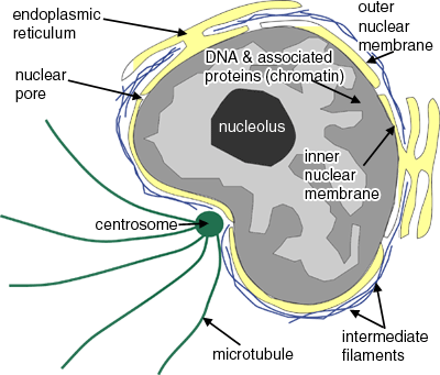
Labeled Diagram Of Cell Membrane : Electron Micrograph The nucleus and mitochondria are two examples. Copy of labeling cell membrane labelled diagram. Some of the major parts of the plasma membrane are : Phospholipid bilayer · phospholipid bilayer ; It supports and helps maintain a cell's shape. 1)cell membrane 2)vacuole 3)nucleus 4)endoplasmic reticulum 5)mitochondria 6)golgi body. Draw a diagram to show the structure of a neuron with myelinated axon ... Draw a diagram to show the structure of a neuron with myelinated axon and label any six parts in it. Class 11. >> Biology. >> Neural Control and Coordination. >> Neuron - The Structural and Functional Unit of Neural System. >> Draw a diagram to show the structure of.
Notes CELL STRUCTURE AND FUNCTION - National Institute … z illustrate the structure of plant and animal cells by drawing labelled diagrams; z describe the structure and functions of plasma membrane, cell wall ... a centrally positioned body which he termed the nucleus . 4.1.2 The cell theory In 1838 M.J. Schleiden and Theodore Schwann formulated the cell theory. ... (As seen in an electron micrograph ...

Nucleus electron micrograph labelled
Histology of the Peripheral Nerves and Light Microscopy The cell body (perykarion) is the dilated region of the neuron that contains a large, euchromatic nucleus with a prominent nucleolus and surrounding perinuclear cytoplasm ... Gamble HJ, Eames RA. An electron microscope study of the connective tissues of human peripheral nerve. J Anat 1964;98:655-663. Salonen V, Roytta M, Peltonen J: The ... Ross willson anatomy and physiology - sr sr - Academia.edu Enter the email address you signed up with and we'll email you a reset link. Histology Laboratory Manual - Columbia University A goblet cell (small intestine, bat) with closely packed mucous droplets (MD). One droplet appears to be close to exiting the cell between two clusters of microvilli (Mv). The apical region of the cell is joined to its neighbor by a zonula occludens (ZO). The nucleus (N) is basal and there is abundant rough endoplasmic reticulum (ER).
Nucleus electron micrograph labelled. Snowflake - Wikipedia A snowflake is a single ice crystal that has achieved a sufficient size, and may have amalgamated with others, which falls through the Earth's atmosphere as snow. Each flake nucleates around a dust particle in supersaturated air masses by attracting supercooled cloud water droplets, which freeze and accrete in crystal form. Complex shapes emerge as the flake … en.wikipedia.org › wiki › Neuroendocrine_tumorNeuroendocrine tumor - Wikipedia Histologically, NETs are an example of "small blue cell tumors," showing uniform cells which have a round to oval stippled nucleus and scant, pink granular cytoplasm. The cells may align variously in islands, glands or sheets. High power examination shows bland cytopathology. Electron microscopy can identify secretory granules. Actin - Wikipedia Actin is a family of globular multi-functional proteins that form microfilaments in the cytoskeleton, and the thin filaments in muscle fibrils.It is found in essentially all eukaryotic cells, where it may be present at a concentration of over 100 μM; its mass is roughly 42 kDa, with a diameter of 4 to 7 nm.. An actin protein is the monomeric subunit of two types of filaments in cells ... Solved Label the transmission electron micrograph of the - Chegg Expert Answer. 100% (23 ratings) Transcribed image text: Label the transmission electron micrograph of the nucleus. Nuclear envelope Nucleolus Nucleus Heterochromatin Reset Zoom.
Solved Please label the electron micrograph to assess your | Chegg.com Question: Please label the electron micrograph to assess your knowledge of the structure and function of a cell's nucleus nuclear pore endoplasma reticulum chromatin nucleolus nuclear envelope This problem has been solved! See the answer Show transcribed image text Expert Answer 100% (3 ratings) 1 ) Nuclear envelo … View the full answer Nucleus - Electron Micrograph - University of Tulsa Slide 5 of 36 Cambridge IGCSE Biology Coursebook (third edition) - Issuu Jun 09, 2014 · The black spots in the electron micrograph in Figure 2.8 are granules of a carbohydrate called glycogen. This is similar to starch. (Starch is never found in animal cells – they store glycogen ... PDF Electron Micrographs (EMs) for laboratories in A215, Basic Human ... - IU There are distinct differences between cilia and microvilli to be seen in electron micrographs: - Cilia are larger (the cilium labeled C about 2.5 microns along its length is probably 5 to 10 microns long); - Cilia contain microtubules, by which they can move.
Electron Micrograph of a Lymphocyte - Netter Images Electron Micrograph of a Lymphocyte. Variant Image ID: 18920. Add to Lightbox. Email this page. Link this page. Print. Please describe! how you will use this image and then you will be able to add this image to your shopping basket. › 40518139 › Ross_willson_anatomyRoss willson anatomy and physiology - sr sr - Academia.edu Enter the email address you signed up with and we'll email you a reset link. Neuroendocrine tumor - Wikipedia Neuroendocrine tumor; Micrograph of a neuroendocrine tumor. HE stain: Specialty: Endocrine oncology : Neuroendocrine tumors (NETs) are neoplasms that arise from cells of the endocrine and nervous systems.They most commonly occur in the intestine, where they are often called carcinoid tumors, but they are also found in the pancreas, lung, and the rest of the body. animal cell electron micrograph labelling Diagram | Quizlet Start studying animal cell electron micrograph labelling. Learn vocabulary, terms, and more with flashcards, games, and other study tools.
File:Anatomy and physiology of animals animal cell electron ...
High-Resolution Scanning Electron Micrograph of the Nucleus of a ... High-Resolution Scanning Electron Micrograph of the Nucleus of a Dividing Cell at Anaphase Variant Image ID: 12976 Add to Lightbox. Save to Lightbox. Email this page; Link this page ; Print; Please describe! how you will use this image and then you will be able to add this image to your shopping basket. Pricing. Price for. Add To Cart ...
Untitled Document
Neuron under Microscope with Labeled Diagram - AnatomyLearner The nucleus is the spherical or elliptical structure in the neuron containing euchromatic staining (pale staining). Again, the shape of the nucleus of a neuron is generally large because of the little cell body cytoplasm. There is a prominent nucleolus evident in the nucleus of a neuron.
DP Topic 1.1 / 1.2 | Biology - Quizizz
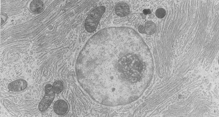
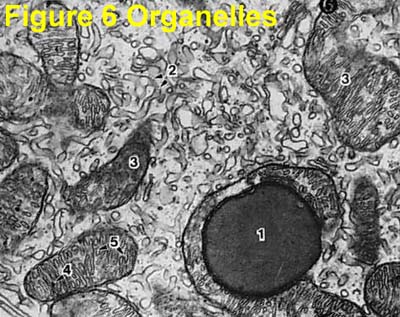
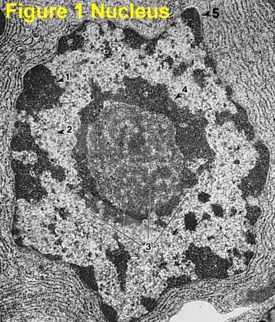
Electron Micrographs of Cell Organelles | Zoology - Biology Discussion This is an electron micrograph of nucleus. (Fig. 17 & 18): (1) Nucleus was discovered by Brown (1831). (2) It is a characteristic entity of almost all eukaryotic cells except mammalian RBCs. (3) The nucleus is generally one but may also be two, four or many.

Nucleus | Celebrate Cytochemistry | Gwen V. Childs, Ph.D.
Animal Cell Electron Microscope Labelled - Q14 Draw a large diagram of ... Using an electron microscope, the electrons can be used to form resolved images of cellular structures of about 3 nm in size. Also provide labels for the different cell structures and organelles. Plant and animal cells have a nucleus inside the cytoplasm. Plant Cells Under Electron Microscope - Micropedia from lh3.googleusercontent.com

Electron Micrograph of Plasma Cells In Connective Tissue
Electron Micrograph of a Cell Nucleus Stock Photo - Image of ... Photo about Electron micrograph of a cell nucleus at high magnification. Image of mitochondria, organelles, cells - 190741290

Cell Micrographs | BioNinja
Defining the ultrastructure of the hematopoietic stem cell ... - eLife Aug 09, 2022 · Hematopoietic stem and progenitor cells (HSPCs) give rise to all blood cell types throughout the life of an organism (Orkin and Zon, 2008).HSPCs reside in a complex microenvironment called the niche that is made up of many different kinds of support cells, including various types of mesenchymal stromal cells (MSCs) and endothelial cells (ECs) …
Solved label the ectron micrograph of an animal cell. | Chegg.com
elifesciences.org › articles › 64835Defining the ultrastructure of the hematopoietic stem cell ... Aug 09, 2022 · Hematopoietic stem and progenitor cells (HSPCs) give rise to all blood cell types throughout the life of an organism (Orkin and Zon, 2008).HSPCs reside in a complex microenvironment called the niche that is made up of many different kinds of support cells, including various types of mesenchymal stromal cells (MSCs) and endothelial cells (ECs) (Pinho and Frenette, 2019).

2.3 Eukaryotic Cells | BioNinja
Nanotwinning-assisted dynamic recrystallization at high ... - Nature May 19, 2022 · Grain refinement is a widely sought-after feature of many metal production processes and frequently involves a process of recrystallization. Some processing methods use very high strain rates and ...

GCE CIE Biology - Animal and Plant Cell Structures and ...
issuu.com › cupeducation › docsCambridge IGCSE Biology Coursebook (third edition) - Issuu Jun 09, 2014 · The black spots in the electron micrograph in Figure 2.8 are granules of a carbohydrate called glycogen. This is similar to starch. (Starch is never found in animal cells – they store glycogen ...

Electron micrographs of SPIO-labeled MSCs. A, Cell nucleus (N ...
animal cell under electron microscope labelled - Be A Terrific Memoir ... Animal Cell Diagram Under Microscope Labeled. Here is an electron micrograph of an animal cell with the labels superimposed. An animal cell represents an eukaryotic cell in which true nucleus and other membrane-bound organelles such as mitochondria Golgi bodies and lysosomes are present. Function cell does in the body.
Electron Micrographs
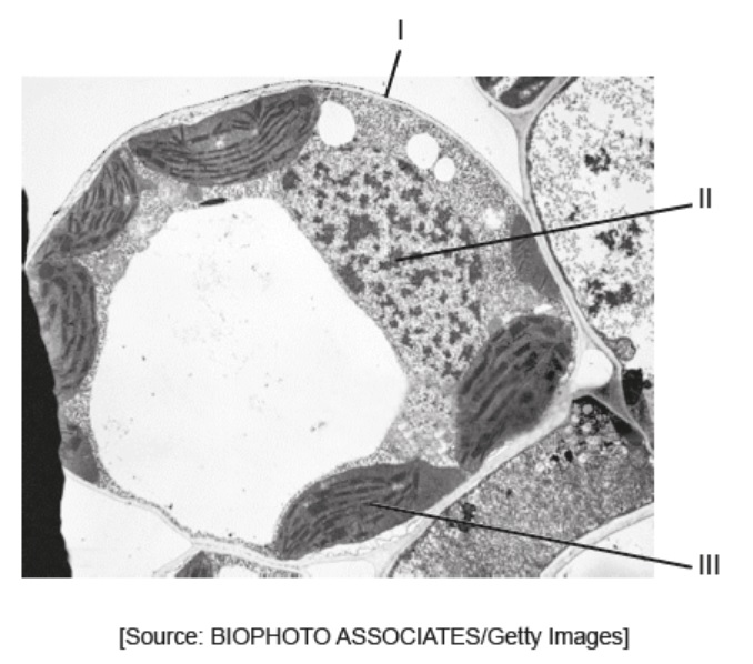
PDF Identifying Organelles from an Electron Micrograph - Ms JMO's Biology ... Courtesy of Dr. Julian Thorpe - EM & FACS Lab, Biological Sciences University Of Sussex The electron micrograph displayed below illustrates many of the plant cell characteristics discussed The cell wall, large central vacuole and chloroplasts are clearly visible Also visible is the clearly defined nucleus containing chromatin
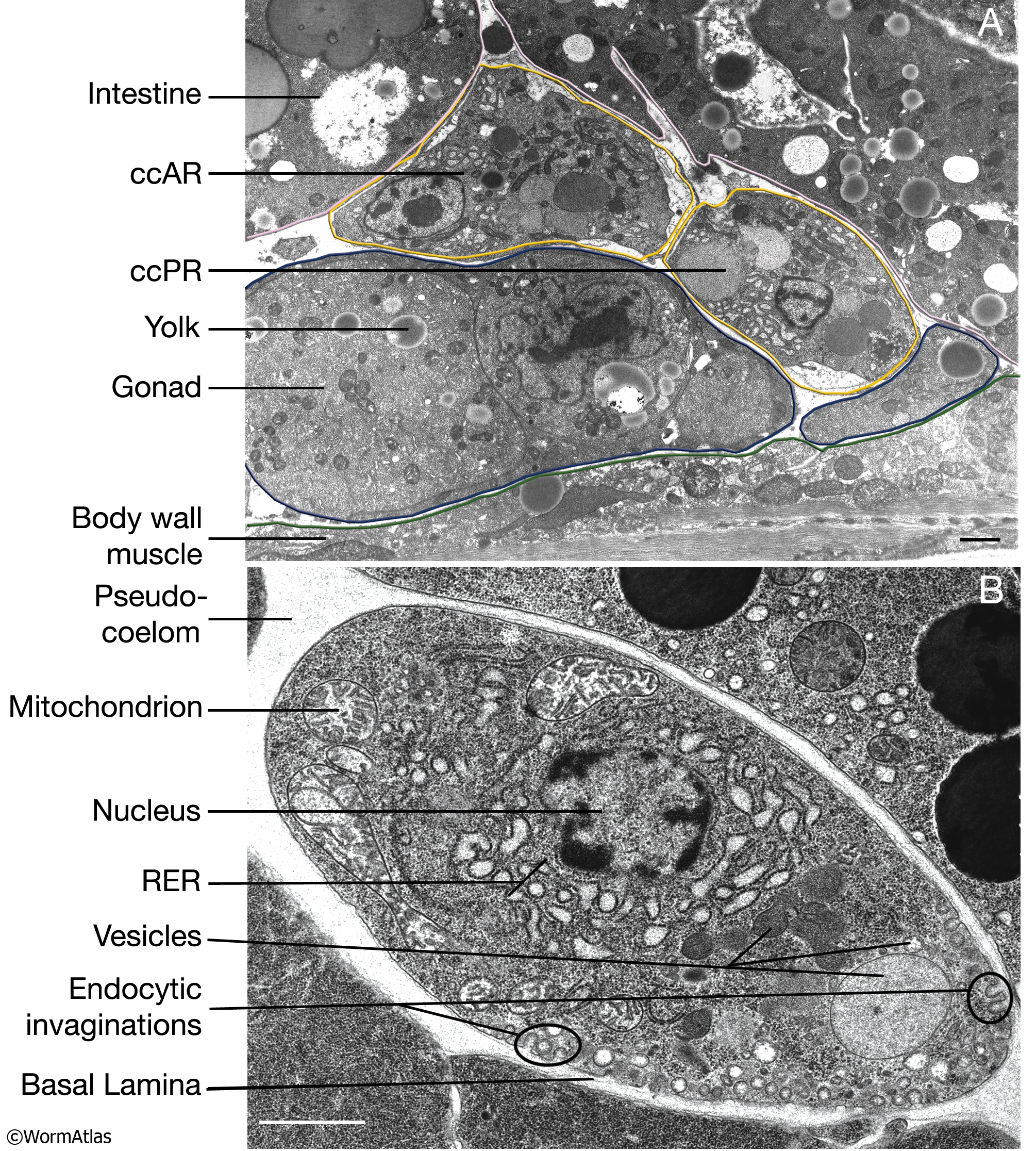
CcFIG 5 Legend
Plant Cell Nucleus Electron Micrograph - Dannie Vanlith Below is a collection of electron micrographs with labelled subcellular structures that you should be able to identify. In mammals it's average diameter is about 6 an electron micrograph of a section through an animal cell nucleus (from an insect cell). In flowering plants, this condition occurs in sieve tube elements.74.

False colour transmission electron microscope (TEM ...
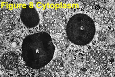
Virtual EM Micrograph List | histology - University of Michigan 021. Plasma Cell: This electron micrograph shows a typical secretory cell, a plasma cell, which secretes immunoglobulin protein. Many of the major types of cellular organelles are visible in this image. In the nucleus, areas of euchromatin and heterochromatin can easily be identified. Virtual Slide.
Electron Micrographs
A, B Electron micrograph of HRP-labeled terminals in the nucleus ... Download scientific diagram | A, B Electron micrograph of HRP-labeled terminals in the nucleus caudalis of the sensory trigeminal nucleus 6 days after vibrissae nerve transection. A Terminal ...

Visualization of the Cell Using EM | Scanning electron ...
Labeling the Cell Flashcards | Quizlet Label the transmission electron micrograph of the nucleus. membrane bound organelles golgi apparatus, mitochondrion, lysosome, peroxisome, rough endoplasmic reticulum nonmembrane bound organelles ribosomes, centrosome, proteasomes cytoskeleton includes microfilaments, intermediate filaments, microtubules Identify the highlighted structures
Electron Micrographs
en.wikipedia.org › wiki › ActinActin - Wikipedia Actin is a family of globular multi-functional proteins that form microfilaments in the cytoskeleton, and the thin filaments in muscle fibrils.It is found in essentially all eukaryotic cells, where it may be present at a concentration of over 100 μM; its mass is roughly 42 kDa, with a diameter of 4 to 7 nm.
Electron Micrograph of Cell Organelles
en.wikipedia.org › wiki › SnowflakeSnowflake - Wikipedia A snowflake is a single ice crystal that has achieved a sufficient size, and may have amalgamated with others, which falls through the Earth's atmosphere as snow. Each flake nucleates around a dust particle in supersaturated air masses by attracting supercooled cloud water droplets, which freeze and accrete in crystal form.
3.3 Eukaryotic Cells – Concepts of Biology – 1st Canadian Edition
Cambridge International AS amp A Level Chemistry Coursebook … Chemists often find it convenient to use a simpler model of the atom in which electrons move around the nucleus in electron shells. Each shell is a certain distance from the nucleus at its own particular energy level (see Section 2.3). ... Draw a labelled diagram to show the structure of a calcium atom. Compare your drawing with that of another ...

Cambridge International AS and A Level Biology Coursebook ...
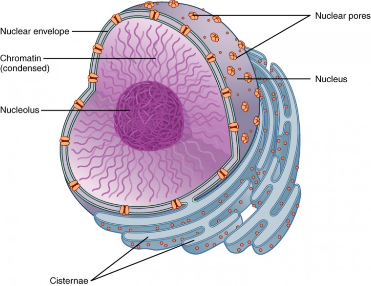
Cell Nucleus - function, structure, and under a microscope The nucleus is a key feature that distinguishes eukaryotic cells, including all animals and plants, from prokaryotic cells (bacteria and archaea). The nucleus (plural: nuclei) stores most of the cell's genetic information in the form of DNA, although mitochondria also contain their own DNA in a very small percentage relative to the nucleus.
Electron Micrographs
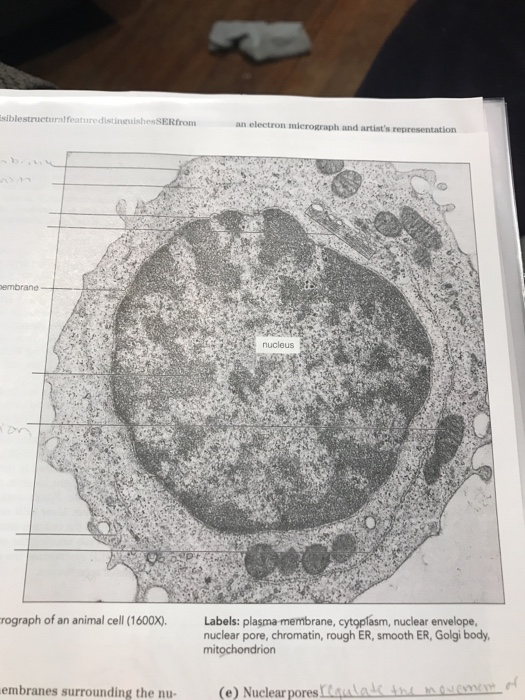
Cell Lab - Yale University The cell's content is divided into two main compartments: the nucleus and the cytoplasm that surrounds the nucleus. Cytoplasm is further divided into organelles, cytosol and inclusions. ... and the electron microscope (magnification up to 500000x). The limit of resolution of the light microscope is 0.2 µm, while the practical limit of ...

Nuclear Envelope | Celebrate Cytochemistry | Gwen V. Childs ...
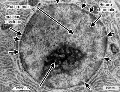
Electron Micrographs - University of Oklahoma Health Sciences Center Figure 1 Micrograph of a nucleus. 1. Heterochromatin 2. Euchromatin 3. Nucleolus 4. Nucleolar associated chromatin 5. Nuclear envelope Figure 2 Micrograph of a portion of a nucleus: What is the round structure (approximately 3 1/2 inches in diameter) seen in the center of this micrograph? 1. Nucleolar associated chromatin 2.
The Nucleus and Cytoplasm | Anatomy and Physiology | | Course ...
› articles › s41563/022/01250-0Nanotwinning-assisted dynamic recrystallization at high ... May 19, 2022 · a, SEM micrograph.b,c, EBSD results: IPF map (b) and KAM map (c), with the inset showing the band-contrast twin map of the marked region.d,e, Bright-field STEM micrographs showing high dislocation ...

1.1 Cell structure | Cells as the basic units of life | Siyavula
Label the transmission electron micrograph of the nucleus. - Transtutors Label the transmission electron micrograph of the nucleus. Expert's Answer Solution.pdf Next Previous Q: Q: Q: Q: Q: Copy And Paste 5 Micrographs With Magnifications That Fall Within The Specified Ranges Into The Text Answer Box. Be Sure To Label Your Images With The Appropriate Name And Magnification. Post Them In The Specified Order. 1.

DP Topic 1.1 / 1.2 | Biology - Quizizz
Nucleus: Definition, Structure, Functions - Biology Learner Oct 14, 2021 · The nucleus is the largest cell organelle in a typical cell. Which is a highly organized globular, ellipsoidal, spherical protoplasmic body. ... Figure: Labelled diagram of Nucleus and its different parts . Nuclear Membrane. ... The electron micrograph and immunocytological techniques show that three distinct regions are observed in the nucleolus.
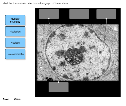
Solved Label the transmission electron micrograph of the ...
Electron Microscope- Definition, Principle, Types, Uses, Labeled Diagram There are two types of electron microscopes, with different operating styles: 1. Transmission Electron Microscope (TEM) The transmission electron microscope is used to view thin specimens through which electrons can pass generating a projection image. The TEM is analogous in many ways to the conventional (compound) light microscope.
The Cell: The Histology Guide
Chapter 20 Scanning Electron Microscopy of Nuclear Structure Scanning Electron Microscopy of the Nucleus and NE. Although the use of a surface imaging technology such as scanning electron microscopy (SEM) might appear an unlikely route to study the nucleus, buried as it is within the cytoplasm, there are many advantages to this type of approach. ... with gold label to mAb 414, an antibody which is ...

2.3.3 Identify structures from electron micrographs of liver ...
Nuclear Labeling | Thermo Fisher Scientific - US Cell-permeant nuclear stains can be used to label nuclei in live cells that have intact, nonpermeable plasma membranes. These dyes will also stain nuclei of cells with compromised membranes such as dead cells or cells that have been fixed and/or permeabilized. Examples of cell-permeant nuclear stains include Hoechst stains and SYTO® stains.

DP Biology: Ultrastructure of cells quiz 1.2
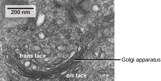
Golgi 5 | Digital Histology Nucleus An electron micrograph of an active fibroblast shows a large Golgi apparatus with its associated vesicles. This cell also shows large accumulations of RER, a feature consistent with a prominent Golgi apparatus. Newly synthesized proteins in the RER are transported to the Golgi by way of transport vesicles. 12,000x
A tour of the cell: View as single page
Histology Laboratory Manual - Columbia University A goblet cell (small intestine, bat) with closely packed mucous droplets (MD). One droplet appears to be close to exiting the cell between two clusters of microvilli (Mv). The apical region of the cell is joined to its neighbor by a zonula occludens (ZO). The nucleus (N) is basal and there is abundant rough endoplasmic reticulum (ER).
Nucleus | Celebrate Cytochemistry | Gwen V. Childs, Ph.D.
Ross willson anatomy and physiology - sr sr - Academia.edu Enter the email address you signed up with and we'll email you a reset link.

Cell Micrographs | BioNinja
Histology of the Peripheral Nerves and Light Microscopy The cell body (perykarion) is the dilated region of the neuron that contains a large, euchromatic nucleus with a prominent nucleolus and surrounding perinuclear cytoplasm ... Gamble HJ, Eames RA. An electron microscope study of the connective tissues of human peripheral nerve. J Anat 1964;98:655-663. Salonen V, Roytta M, Peltonen J: The ...
Biology, The Cell, Cell Structure, The Endomembrane System ...

Electron micrograph of pancreatic exocrine cells from control ...
Electron Micrographs

1.1 Cell structure | Cells as the basic units of life | Siyavula
The Cell: The Histology Guide

A Transmission electron micrograph (TEM) of a transverse ...

The Nucleus - Cell Organelles Ep 1 - Zoë Huggett Tutorials
The Cell: The Histology Guide
Electron Micrographs

AICE Biology Chapter 1: Animal Cell Electron Micrograph ...
Post a Comment for "41 nucleus electron micrograph labelled"