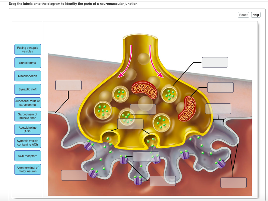
40 drag the labels onto the diagram to identify the components of a cholinergic synapse.
Synapse Activity Teaching Resources | Teachers Pay Teachers NERVE CELLS AND SYNAPSES DIAGRAM WORKSHEETIncluded in this resource:Nerve Cell Diagram Worksheet - Looking at parts of the nerve cell, students can label and describe the functions (e.g. nucleus, axon, dendrites, myelin sheath etc). Students are also asked to define what a neuron is and the three types or neuron, linked to a simple diagram. Answered: Would drugs designed to disrupt cell… | bartleby 11th Edition. ISBN: 9781305251052. Author: Michael Cummings. Publisher: Cengage Learning. expand_less. 1 A Perspective On Human Genetics 2 Cells And Cell Division 3 Transmission Of Genes From Generation To Generation 4 Pedigree Analysis In Human Genetics 5 The Inheritance Of Complex Traits 6 Cytogenetics: Karyotypes And Chromosome Aberrations 7 ...
A&P2 Lab 1 HW Flashcards | Quizlet Drag the labels onto the diagram to identify the parts of a myelinated PNS neuron. look at pic. Drag the labels to identify the structural components of a typical synapse. look at pic. Rabies illustrates a negative consequence to otherwise healthy retrograde flow within axons.
Drag the labels onto the diagram to identify the components of a cholinergic synapse.
Structure of Chemical Synapses | SynapseWeb Structure of Chemical Synapses. Functional communication between neurons occurs at specialized junctions called synapses. The most common types of synapses in the brain use chemicals (more specifically, neurotransmitters) to communicate between neurons. These are called chemical synapses. A presynaptic element, an axon, and a postsynaptic ... Central nervous system - Wikipedia Central nervous system. The central nervous system ( CNS) is the part of the nervous system consisting primarily of the brain and spinal cord. The CNS is so named because the brain integrates the received information and coordinates and influences the activity of all parts of the bodies of bilaterally symmetric and triploblastic animals —that ... A & P Ch 12 & Ch 18 Flashcards | Quizlet Drag the labels to identify the sequence of events that occurs at a synapse. 1. an action potential arrives at the synaptic terminal 2. calcium channels open, and calcium ions enter the synaptic terminal
Drag the labels onto the diagram to identify the components of a cholinergic synapse.. Chapter 07 Homework Answers.pdf - Course Hero Part A - Synaptic response to an action potential Drag the labels to identify the sequence of events that occurs at a synapse. ANSWER: Help Reset Sensory Somatic Motor Autonomic 10/19/2017 Chapter 07 Homework Correct Chapter 7 Chapter Test Question 4 Part A What part of a neuron receives signals and sends a message to the cell body? Solved (Neuror/ Nervous Tissue Homework Adaptive Follow-Up - Chegg Anatomy and Physiology. Anatomy and Physiology questions and answers. (Neuror/ Nervous Tissue Homework Adaptive Follow-Up Art-labeling Activity: Components of a cholinergic synapse - Part A Drag the labels onto the diagram to identify the components of a cholinergic synapse. Reset Help Acetylcholine Acetyl-CoA cal Postsynapsc membrane Acetylcholine receptor Mitochondrion Calcium on Synaptic de 60 (ACE) Acetylcholinesterase. Neuron Action Potential Sequence of Events - GetBodySmart An action potential occurs when a portion of the membrane rapidly depolarizes and then repolarizes again to the original resting state. The process is initiated by a threshold level stimulus, such as a nearby change in membrane potential (threshold potential, local potential). At threhsold (about -55mV), many Na+ voltage-gated channels open ... Neuromuscular Junctions and Muscle Contractions - Course Hero Neuromuscular Junctions. Skeletal muscle cell contraction occurs after a release of calcium ions from internal stores, which is initiated by a neural signal. Each skeletal muscle fiber is controlled by a motor neuron, which conducts signals from the brain or spinal cord to the muscle. The following list presents an overview of the sequence of ...
Chemical level of organization Flashcards | Quizlet Drag the labels onto the diagram to identify the steps in a reaction both with and without enzymes. Click card to see definition 👆. Tap card to see definition 👆. ... Click again to see term 👆. Tap again to see term 👆. Drag the labels onto the diagram to identify the various components of the pH scale. Click card to see definition 👆. ReaderUi ReaderUi synapse labeling quiz - vt6rentals.com synapse labeling quiz women's mma atomweight rankings. Menu. bellator dublin 2022 tickets; big bear homes for sale zillow Synapses | Anatomy and Physiology I | | Course Hero The term synapse means "coming together." Where two structures or entities come together, they form a synapse. Although one can use the word synapse to mean any cellular junction, in physiology we traditionally limit its usage to: the junction of two neurons, the junction between a neuron and a target cell (ex. the neuromuscular junction), or the interface between adjacent cardiac muscle ...
Solved Drag the labels onto the diagram to identify the - Chegg Expert Answer. Visceral motor nuclei in hypothalamus. Smooth muscle. G …. View the full answer. Transcribed image text: Prag the labels onto the diagram to identify the components of the autonomic nervous system! Reset Help Cardiac muscle Smooth muscle Brain Ganglionic neurons Preganglionic neuron Visceral Effectors Adipocytes Autonomic ... Divisions of the Autonomic Nervous System - Lumen Learning The terms cholinergic and adrenergic refer not only to the signaling molecule that is released but also to the class of receptors that each binds. The cholinergic system includes two classes of receptor: the nicotinic receptor and the muscarinic receptor. Both receptor types bind to ACh and cause changes in the target cell. Human Anatomy & Physiology Laboratory Manual Main Version 10th Edition ... Identifying the Parts of a Microscope 1. Using the proper transport technique, obtain a microscope and bring it to the laboratory bench. • Record the number of your microscope in the Summary Chart (page 31). Compare your microscope with the photograph (Figure 3.1) and identify the following microscope parts: Base: Supports the microscope. Synapse: Definition, Mechanism and Properties (With Diagram) ADVERTISEMENTS: In this article we will discuss about:- 1. Definition of Synapse 2. Mechanism of Synaptic Transmission 3. Properties. Definition of Synapse: Synapse can be defined as functional junction between parts of two different neurons. There is no anatomical continuity between two neurons involved in the formation of synapse. At level of synapse, impulse gets […]
Mastering A and P Assignment Unit 2 - Nervous System - StuDocu Drag the terms on the left to the appropriate blanks on the right to complete the sentences. Hint 1. Cholinergic receptors. Cholinergic receptors are classified by the type of agonist that can activate them. Hint 2. Adrenergic receptors. The adrenergic receptors are classified either as alpha-adrenergic or beta-adrenergic.
NEURON STRUCTURE AND CLASSIFICATION - Brigham Young University-Idaho Structural classification of neurons. 1) Bipolar; 2) Multipolar and 3) Unipolar. Bipolar neurons have only two processes that extend in opposite directions from the cell body. One process is called a dendrite, and another process is called the axon. Although rare, these are found in the retina of the eye and the olfactory system.
Chpt. 1, 3, 24, and 9 HW - Weebly Drag each image to the appropriate location in the sequence. High Blood glucose levels Blood glucose becomes high --> Pancreas releases insulin -->Insulin binds to receptors on target cells --> cells take in glucose --> Blood glucose returns to normal 6. Drag the appropriate labels to their respective targets. (from left to right)
BIO Flashcards - Quizlet Study with Quizlet and memorize flashcards containing terms like Drag the labels onto the diagram to identify the components of replicating DNA strands., Drag the labels onto the diagram to identify the stages in which the lagging strand is synthesized., Drag the correct labels onto the diagram to identify the structures and molecules involved in translation. and more.
16.4 The Peripheral Nervous System - Concepts of Biology - 1st Canadian ... Describe the organization and function of the sensory-somatic nervous system. The peripheral nervous system (PNS) is the connection between the central nervous system and the rest of the body. The CNS is like the power plant of the nervous system. It creates the signals that control the functions of the body.
Chapter 11 Homework Flashcards - Quizlet Drag the labels to identify the sequence of events that occurs at a synapse. Drag the labels onto the diagram to identify the various synapse structures. Arrange the parts in order, from left to right, of a successful direct depolarization path within one neuron.
Structure of the Autonomic Nervous System - Course Hero The ANS is unique in that it requires a sequential two-neuron efferent pathway; the preganglionic neuron must first cross a synapse onto a postganglionic neuron before innervating the target organ. The preganglionic, or first neuron will begin at the outflow and will cross a synapse at the postganglionic, or second neuron's cell body.
Myelinated axons with the largest diameter Part A Drag the labels onto the diagram to identify the structural components at a cholinergic synapse when an action potential arrives at the synaptic knob.
A & P Ch 12 & Ch 18 Flashcards | Quizlet Drag the labels to identify the sequence of events that occurs at a synapse. 1. an action potential arrives at the synaptic terminal 2. calcium channels open, and calcium ions enter the synaptic terminal
Central nervous system - Wikipedia Central nervous system. The central nervous system ( CNS) is the part of the nervous system consisting primarily of the brain and spinal cord. The CNS is so named because the brain integrates the received information and coordinates and influences the activity of all parts of the bodies of bilaterally symmetric and triploblastic animals —that ...
Structure of Chemical Synapses | SynapseWeb Structure of Chemical Synapses. Functional communication between neurons occurs at specialized junctions called synapses. The most common types of synapses in the brain use chemicals (more specifically, neurotransmitters) to communicate between neurons. These are called chemical synapses. A presynaptic element, an axon, and a postsynaptic ...











Post a Comment for "40 drag the labels onto the diagram to identify the components of a cholinergic synapse."