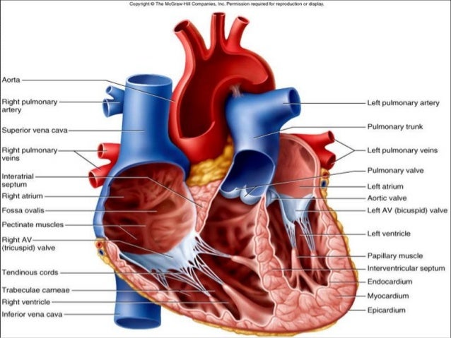
43 correctly label the internal anatomy of the heart
Human Heart (Anatomy): Diagram, Function, Chambers, Location in ... - WebMD The heart is a muscular organ about the size of a fist, located just behind and slightly left of the breastbone. The heart pumps blood through the network of arteries and veins called the... Heart Anatomy: Labeled Diagram, Structures, Blood Flow ... - EZmed Let's begin with the chambers of the heart. There are 4 chambers, labeled 1-4 on the diagram below. To help simplify things, we can convert the heart into a square. We will then divide that square into 4 different boxes which will represent the 4 chambers of the heart.
Heart: Anatomy and Function - Cleveland Clinic The heart is a fist-sized organ that pumps blood throughout your body. It's the primary organ of your circulatory system. Your heart contains four main sections (chambers) made of muscle and powered by electrical impulses. Your brain and nervous system direct your heart's function. Cleveland Clinic is a non-profit academic medical center.

Correctly label the internal anatomy of the heart
Correctly Label The Following Internal Anatomy Of The Heart When you study the anatomy of the heart, you will see that it has three main anatomical features. Among them are the aorta, the vena cava, and the pulmonary veins. The heart is made of tissue. It needs nutrients and oxygen. The chambers of the heart are filled with blood. However, the heart does not receive nourishment from the blood. Diagram of Human Heart and Blood Circulation in It Four Chambers of the Heart and Blood Circulation. The shape of the human heart is like an upside-down pear, weighing between 7-15 ounces, and is little larger than the size of the fist. It is located between the lungs, in the middle of the chest, behind and slightly to the left of the breast bone. The heart, one of the most significant organs ... A Labeled Diagram of the Human Heart You Really Need to See The blood pumped by the heart not only provides nutrients to the body cells, but also removes the waste materials from different parts of the body. The above labeled diagram can be modified as per your requirements for kids. Heart diagram for kids can be printed out and colored, to make it easier to understand.
Correctly label the internal anatomy of the heart. Layers of the heart: Epicardium, myocardium, endocardium - Kenhub It lines the inner surfaces of the heart chambers, including the heart valves. The endocardium has two layers. The inner layer lines the heart chambers and is made of endothelial cells. Superiorly, is the second layer: a subendocardial connective tissue which is continuous with the connective tissue of the myocardium. Chapter 20-Cardiovascular System Flashcards - Quizlet Correctly label the following internal anatomy of the heart. b Place the labels in order denoting the flow of oxygenated blood through the heart beginning with the vessels that bring blood back to the heart from the lungs. Correctly label the following coronary blood vessels of the heart. Human Heart - Anatomy, Functions and Facts about Heart Practice your understanding of the heart structure. Drag and drop the correct labels to the boxes with the matching, highlighted structures. Instructions to use: Hover the mouse over one of the empty boxes. One part in the image gets highlighted. Identify the highlighted part and drag and drop the correct label into the same box. Label the heart - Science Learning Hub In this interactive, you can label parts of the human heart. Drag and drop the text labels onto the boxes next to the heart diagram. If you want to redo an answer, click on the box and the answer will go back to the top so you can move it to another box. If you want to check your answers, use the Reset Incorrect button.
Ch. 19 Circulatory System- heart Flashcards - Quizlet Correctly label the internal anatomy of the heart. Correctly label the following internal anatomy of the heart. Drag each label to the location of each structure described. Explanation The heart functions to first pump deoxygenated blood returning from the body to the lungs in order to release carbon dioxide and reoxygenate the blood. Heart Anatomy | Anatomy and Physiology | | Course Hero Describe the internal and external anatomy of the heart Identify the tissue layers of the heart Relate the structure of the heart to its function as a pump Compare systemic circulation to pulmonary circulation Identify the veins and arteries of the coronary circulation system The Heart | Circulatory Anatomy - Visible Body The epicardium covers the heart, wraps around the roots of the great blood vessels, and adheres the heart wall to a protective sac. The middle layer is the myocardium. This strong muscle tissue powers the heart's pumping action. The innermost layer, the endocardium, lines the interior structures of the heart. 2. Anatomy and Function of the Heart's Electrical System The electrical stimulus travels down through the conduction pathways and causes the heart's ventricles to contract and pump out blood. The 2 upper chambers of the heart (atria) are stimulated first and contract for a short period of time before the 2 lower chambers of the heart (ventricles). The electrical impulse travels from the sinus node to ...
[Solved] Check my work Correctly label the followiting Internal anatomy ... e correcty label the following internal anatomy of the heart left atrium interventricular septum papillary muscles right ventricle opening of superior left ventricle vena cava fossa ovalis right atrium right atrium pectinate tricuspid valve muscles right ventricle rabeculae camae papillary muscle o download attachments: attachment1.png skip … Diagrams, quizzes and worksheets of the heart - Kenhub Using our unlabeled heart diagrams, you can challenge yourself to identify the individual parts of the heart as indicated by the arrows and fill-in-the-blank spaces. This exercise will help you to identify your weak spots, so you'll know which heart structures you need to spend more time studying with our heart quizzes. correctly label the following internal anatomy of the heart correctly label the following internal anatomy of the heart The human heart has three chambers, which are also known as the atria, the ventricles, and the atrioventricular (A-V) valves. Heart Anatomy Labeling Game - PurposeGames.com About this Quiz This is an online quiz called Heart Anatomy Labeling Game There is a printable worksheet available for download here so you can take the quiz with pen and paper. Your Skills & Rank Total Points 0 Get started! Today's Rank -- 0 Today 's Points One of us! Game Points 19 You need to get 100% to score the 19 points available Actions
Human Heart - Diagram and Anatomy of the Heart - Innerbody Because the heart points to the left, about 2/3 of the heart's mass is found on the left side of the body and the other 1/3 is on the right. Anatomy of the Heart Pericardium. The heart sits within a fluid-filled cavity called the pericardial cavity. The walls and lining of the pericardial cavity are a special membrane known as the pericardium.
Correctly label the following internal anatomy of the heart. Right ... Correctly label the following internal anatomy of the heart. Right pulmonary artery Pulmonary trunk Right pulmonary veins Pulmonary valve Right ventricle Aorta Left pulmonary veins Left pulmonary artery Right atrium Apr 01 2022 05:38 PM Expert's Answer Solution.pdf Next Previous
Solved Correctly label the following parts of the internal - Chegg Correctly label the following parts of the internal anatomy of the heart. Right pulmonary veins Aorta Left pulmonary veins Pulmonary semilunar Left ventricle Right ventricle Bicuspid valve Tricuspid valve Right pulmonary artery Septum Pulmonary trunk Right atrium Left pulmonary artery Inferior vena cava Left atrium Superior vena cava Reset Zoom
19.1 Heart Anatomy - Anatomy and Physiology 2e | OpenStax Location of the Heart. The human heart is located within the thoracic cavity, medially between the lungs in the space known as the mediastinum. Figure 19.2 shows the position of the heart within the thoracic cavity. Within the mediastinum, the heart is separated from the other mediastinal structures by a tough membrane known as the pericardium, or pericardial sac, and sits in its own space ...
Human Heart Diagram Labeled - Science Trends Anatomy Of The Heart. The human heart usually weighs somewhere between 10 to 12 ounces in men and between 8 to 10 ounces in women, and in terms of size is roughly the size of the fist. The heart has four different chambers: the left and right ventricles and the left and right atriums. The chambers of the heart and the valves that regulate blood ...
Correctly label the following internal anatomy of the heart. Fossa ... Correctly label the following parts of the internal anatomy of the heart. Place your cursor over the boxes for more information papillary muscles bicuspid valve right atrium septum pulmonary semilunar valve eft atrium chordae tendineae pulmonary...
The Anatomy of the Heart, Its Structures, and Functions The heart is the organ that helps supply blood and oxygen to all parts of the body. It is divided by a partition (or septum) into two halves. The halves are, in turn, divided into four chambers. The heart is situated within the chest cavity and surrounded by a fluid-filled sac called the pericardium. This amazing muscle produces electrical ...
Heart Labeling Quiz: How Much You Know About Heart Labeling? Here is a Heart labeling quiz for you. The human heart is a vital organ for every human. The more healthy your heart is, the longer the chances you have of surviving, so you better take care of it. Take the following quiz to know how much you know about your heart. Questions and Answers. 1.


Post a Comment for "43 correctly label the internal anatomy of the heart"